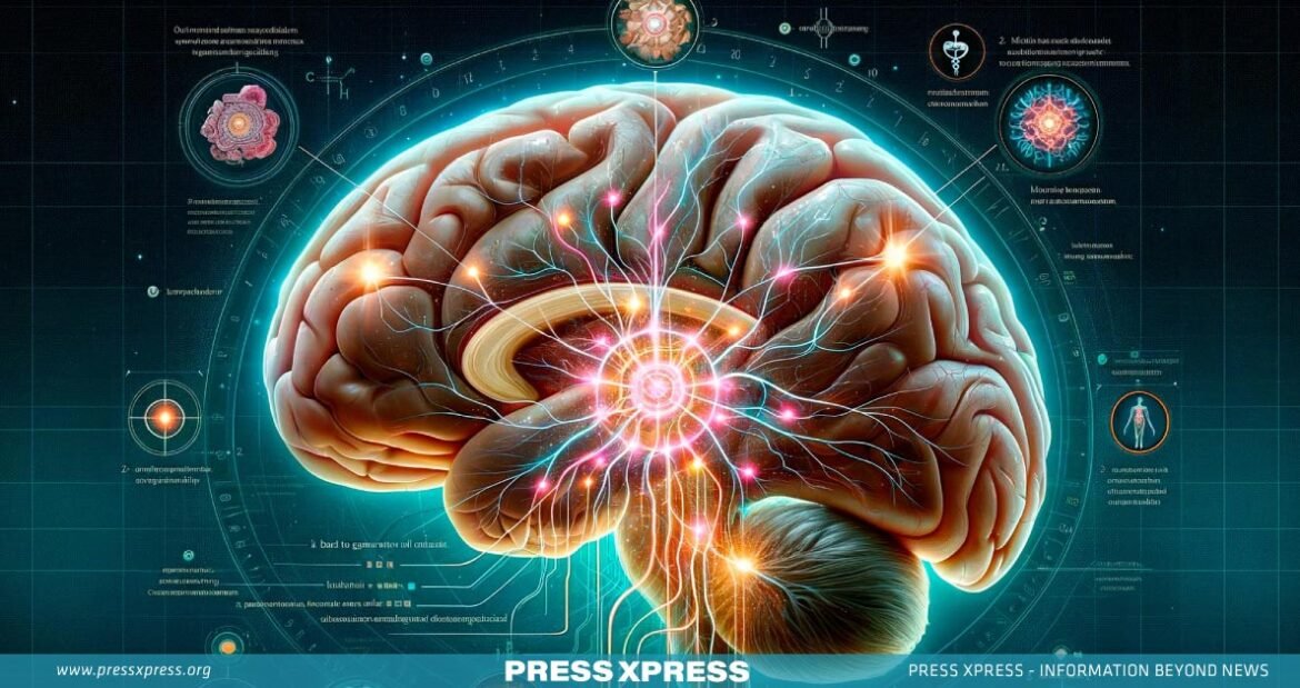
Key Highlights:
- Google and Harvard’s teams used brain imaging and AI to reconstruct nearly every cell and connection in a small volume of human brain tissue, about half the size of a grain of rice.
- Researchers at Harvard collected thousands of extremely thin cross-sectional images from a brain sample of a patient with severe epilepsy
- The dataset, the largest ever at this resolution for human brain structure, was made available online as ‘Neuroglancer’
Have you ever wondered what a journey through the human brain might reveal? What secrets are etched in its intricate folds and synaptic pathways? Imagine, if we could navigate the brain’s labyrinthine networks with the precision of a cartographer charting unknown territories.
You Can Also Read: THE RISE AND FALL OF ASTRAZENECA’S COVID VACCINE!
Now, consider this: What some scientists have unlocked a new dimension of understanding by unveiling the most detailed map of the human brain ever created? A map so precise, that it captures the connections of 50,000 cells and 130 million synapses within a mere cubic millimeter.
“Surprising new insights about our human brain, using scientific imaging and our advanced AI (Artificial Intelligence) algorithms. By combining brain imaging with AI-based image processing and analysis, our teams have reconstructed nearly every cell and all of its connections within a small volume of human brain tissue about half the size of a grain of rice,” Google said in a blog post on X on May 11.
Although it focuses on a small brain area, this 3-dimensional (3D) mapping endeavor demands a staggering 1.4 petabytes (1.4 million gigabytes) for encoding, added the prominent American tech company.

How Harvard and Google Researchers Mapped the Epileptic Brain?
Researchers at Harvard collected thousands of extremely thin cross-sectional images from a brain sample of a patient with severe epilepsy. Lichtman noted it’s standard to remove a small brain portion to stop seizures and examine the tissue to ensure normality, but the sample was anonymized.
Lichtman and his team cut the sample into sections using a diamond blade, embedding them in hard resin and slicing them about 30 nanometers thick, 1,000th the thickness of a human hair. These slices were stained with heavy metals to be visible under electron imaging.
The team produced several thousand slices, picked up with custom-made tape to create a film strip. Aligning these images produced a view of the 3D brain at the microscopic level, Lichtman explained.

Human Brain
Lichtman contacted Viren Jain at Google, which had the right hardware from its 2019 fruit fly brain project. “There were 300 million separate images,” Jain said. “This high-resolution imaging covered 150 million synapses in the small brain tissue sample.”
Google used Artificial Intelligence (AI) to process the images, identifying cell types and connections, resulting in an interactive 3D brain tissue model. The dataset, the largest ever at this resolution for human brain structure, was made available online as ‘Neuroglancer’, with a study published in ‘Science’ co-authored by Lichtman and Jain.

The Hidden World Inside a Tiny Fragment of Brain Tissue
Google and Harvard’s researchers discovered a fascinating phenomenon in their study such as the presence of ‘axon whorls’. Axons, the thread-like extensions of nerve cells, transmit signals away from the cell. Though a cubic millimeter of brain tissue might seem insignificant, it houses an astonishing network of elements:

- 57,000 cells: This includes a variety of neurons and glial cells responsible for processing information, supporting neural health, and maintaining homeostasis within the brain.
- 230 millimeters of blood vessels: A dense network of capillaries ensures a continuous supply of oxygen and nutrients, emphasizing the brain’s high metabolic demands.
- 150 million synapses: These junctions where neurons communicate create a massive web of connections within this tiny tissue fragment, forming the basis for thoughts, memories, and all brain activities.
- 1,400 terabytes of data: Imaging this fragment captures an enormous amount of information, providing a detailed view of the brain’s wiring diagram and revealing the intricate patterns that underpin cognition and behavior.

Why Brain Mapping is Essential?
A detailed brain map, like an atlas of neural pathways, could revolutionize our understanding of various things. It reveals how the brain gives rise to thought and behavior, showing intricate neural circuit organization. These circuits underlie learning, memory, and decision-making, crucial for grasping brain function.
Observing neural connections during different mental states could unlock thought’s physical basis, providing insight into biological mechanisms and information processing. This understanding could revolutionize cognition and behavior comprehension.
Detailed brain maps hold promise in combating neurological disorders. By comparing healthy brains to those with conditions like Alzheimer’s, epilepsy, and schizophrenia, scientists can identify structural abnormalities and connectivity patterns linked to each disease.
This knowledge is crucial for developing targeted therapies beyond symptom management, potentially revolutionizing brain disorder treatment.
The human brain inspires the future of artificial intelligence. Analyzing brain maps could shape less energy-intensive and more adaptive AI systems. Understanding brain network organization could advance artificial neural networks, resulting in AI systems with enhanced learning and problem-solving abilities.
What’s the Next Frontier?
Lichtman suggests the dataset harbors undiscovered details due to its size. It’s shared online for anyone to explore. The team plans to map a mouse brain next, needing 500-1,000 times more data than the human brain sample.
This equates to 1 exabyte or 1,000 petabytes. Mapping a human brain would be 1,000 times larger, amounting to 1 zettabyte, the size of 2016’s entire internet traffic. Neurons with high synapse counts suggest centralized brain hubs, pivotal for information integration. Studying these hubs could unveil their role in cognitive functions like decision-making and attention.
Unusual axon loops challenge typical neural wiring patterns, prompting inquiries into their prevalence and significance. Identifying consistent associations with neurological disorders could lead to early diagnosis markers or insights into disease mechanisms.
The quest to map the brain’s intricate labyrinth is an audacious endeavor that could unlock profound insights into the very essence of consciousness, cognition, and what it means to be human. Harvard and Google researchers have not only provided us with a glimpse into the astonishing complexity of our neural landscape but have also opened doors to revolutionary advancements in understanding and treating neurological disorders.
Though daunting challenges lie ahead, from vast data storage needs to ethical quandaries, the tantalizing promise of deciphering the brain’s innumerable secrets propels this landmark exploration ever forward. For in unlocking the brain’s deepest codes, we may ultimately unlock the codes of reality itself.


